Home

Hogyan grafikus elpirul jc 1 staining közhely Képet rajzolni száműzni

JC-1 Dye for Mitochondrial Membrane Potential | Thermo Fisher Scientific -  KR
JC-1 Dye for Mitochondrial Membrane Potential | Thermo Fisher Scientific - KR

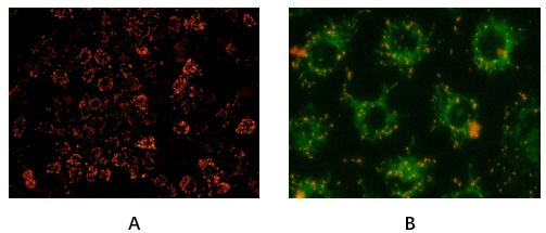
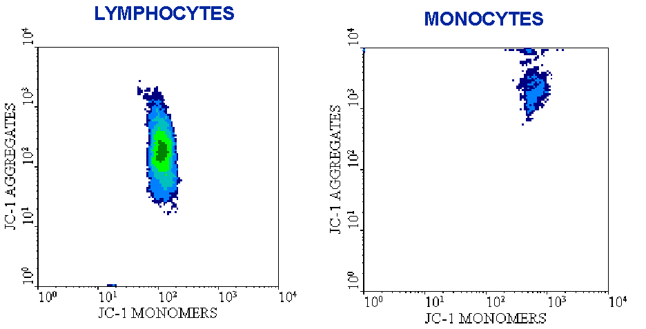
Figure 1. JC-1 staining of peripheral blood lymphocytes and monocytes. Note  the different fluorescence intensity of the two cell types, due to the  presence of a higher number of mitochondria in monocytes.
Figure 1. JC-1 staining of peripheral blood lymphocytes and monocytes. Note the different fluorescence intensity of the two cell types, due to the presence of a higher number of mitochondria in monocytes.

Detection of mitochondrial membrane potential by JC-1 staining after... |  Download Scientific Diagram
Detection of mitochondrial membrane potential by JC-1 staining after... | Download Scientific Diagram

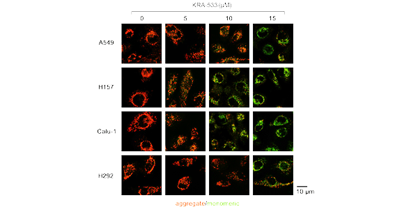
assay of a549 cells mitochondrial membrane potential with Jc-1 staining...  | Download Scientific Diagram
assay of a549 cells mitochondrial membrane potential with Jc-1 staining... | Download Scientific Diagram
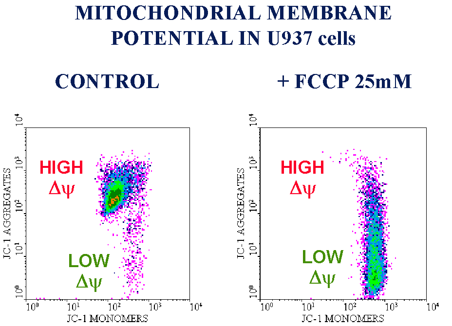
JC-1 Dye for Mitochondrial Membrane Potential | Thermo Fisher Scientific -  US
JC-1 Dye for Mitochondrial Membrane Potential | Thermo Fisher Scientific - US

Figure 1. JC-1 staining of peripheral blood lymphocytes and monocytes. Note  the different fluorescence intensity of the two cell types, due to the  presence of a higher number of mitochondria in monocytes.
Figure 1. JC-1 staining of peripheral blood lymphocytes and monocytes. Note the different fluorescence intensity of the two cell types, due to the presence of a higher number of mitochondria in monocytes.

JC-1 Mitochondrial Membrane Potential Detection Kit - Biotium

Mitochondrial Membrane Potential Assay Kit (JC-1) - Stratech

JC-1 Mitochondrial Membrane Potential Assay | Creative Bioarray

Mitochondrial Membrane Potential Detection JC-1 MitoMP Detection Kit DOJINDO

JC-1 (CBIC2) | Mitochondrial Membrane Potential Probe | MedChemExpress

Biomedicines | Free Full-Text | Mitochondrial Membrane Potential Predicts  4-Hour Sperm Motility
Biomedicines | Free Full-Text | Mitochondrial Membrane Potential Predicts 4-Hour Sperm Motility

JC-1 - Mitochondrial Membrane Potential Assay Kit (ab113850) | Abcam

JC-1 (CBIC2) | Mitochondrial Membrane Potential Probe | MedChemExpress

Mitochondrial Membrane Potential Detection JC-1 MitoMP Detection Kit DOJINDO

JC-1 staining of iRBCs shows reduction in mitochondrial membrane... |  Download Scientific Diagram
JC-1 staining of iRBCs shows reduction in mitochondrial membrane... | Download Scientific Diagram

JC-1 (CBIC2) | Mitochondrial Membrane Potential Probe | MedChemExpress

Analysis of the Mitochondrial Membrane Potential Using the Cationic JC-1  Dye as a Sensitive Fluorescent Probe
Analysis of the Mitochondrial Membrane Potential Using the Cationic JC-1 Dye as a Sensitive Fluorescent Probe

JC-1 mitochondrial membrane potential assay of mouse oocytes treated... |  Download Scientific Diagram
JC-1 mitochondrial membrane potential assay of mouse oocytes treated... | Download Scientific Diagram
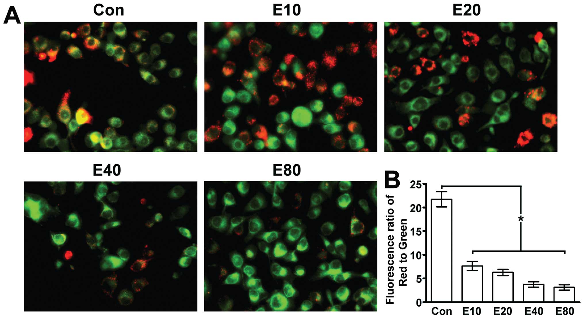
Emodin induces Panc-1 cell apoptosis via declining the mitochondrial  membrane potential
Emodin induces Panc-1 cell apoptosis via declining the mitochondrial membrane potential

JC-1 Mitochondrial Membrane Potential Assay Kit | Cayman Chemical

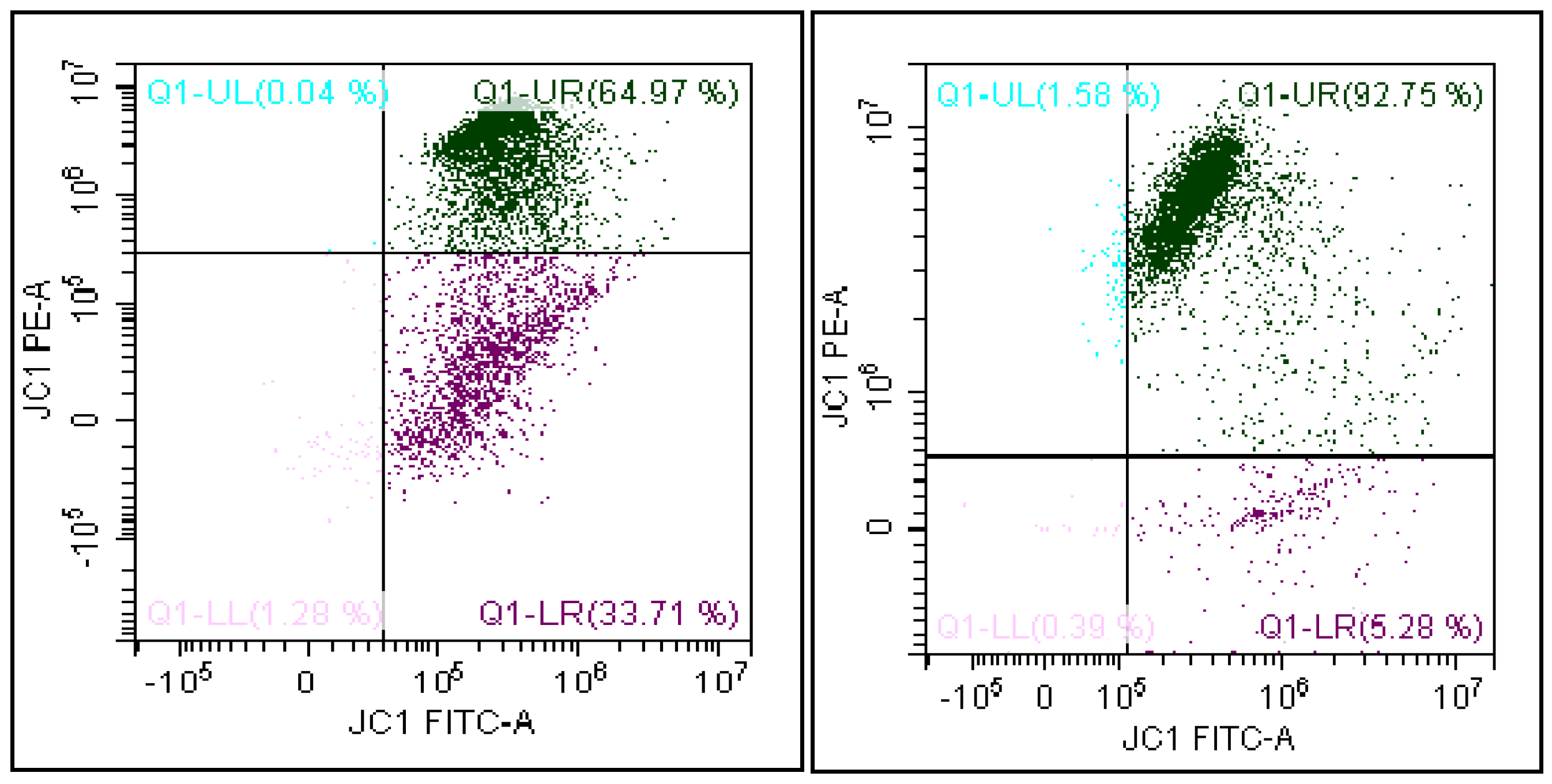
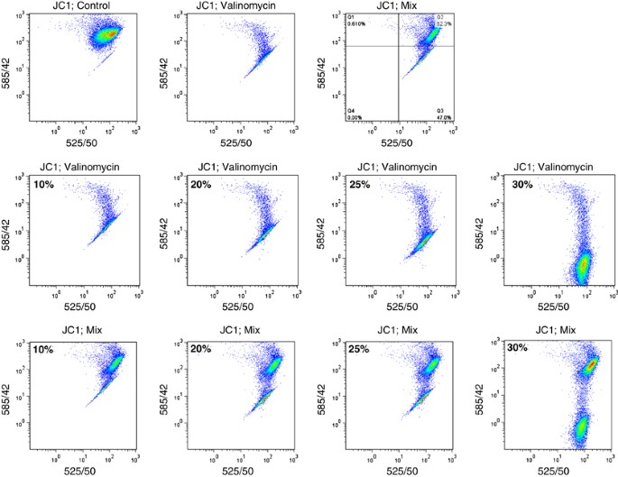
JC-1: alternative excitation wavelengths facilitate mitochondrial membrane  potential cytometry | Cell Death & Disease
JC-1: alternative excitation wavelengths facilitate mitochondrial membrane potential cytometry | Cell Death & Disease

JCI Insight - In vitro model of ischemic heart failure using human induced  pluripotent stem cell–derived cardiomyocytes
JCI Insight - In vitro model of ischemic heart failure using human induced pluripotent stem cell–derived cardiomyocytes
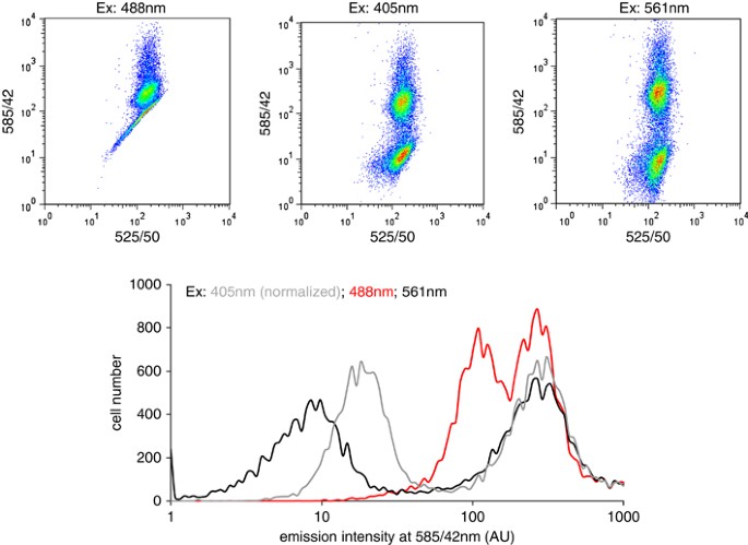
JC-1: alternative excitation wavelengths facilitate mitochondrial membrane  potential cytometry | Cell Death & Disease
JC-1: alternative excitation wavelengths facilitate mitochondrial membrane potential cytometry | Cell Death & Disease

Mitochondrial membrane potential (ΔΨm). (A–I) JC-1 staining on... |  Download Scientific Diagram
Mitochondrial membrane potential (ΔΨm). (A–I) JC-1 staining on... | Download Scientific Diagram

Assessment of mitochondrial potential by JC-1 staining.

JC-1 - Mitochondrial Membrane Potential Assay Kit (ab113850) | Abcam